The Eye
23 January 2026 2026-01-29 14:09The Eye
Myopia
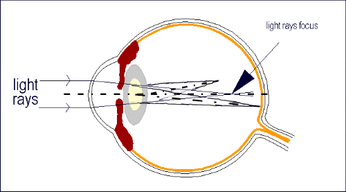
In myopia the distance between the cornea and the retina is greater than normal. A parallel beam that enters the eye does not focus on the retina, but in front of him.

Those with myopia see near objects clearly but far away objects appear blurred. With myopia, the eyeball is too long, or the cornea is too steep, so images are focused in the vitreous inside the eye rather than on the retina at the back of the eye.
The continuous increase during intramedullary axis of the eye during development is responsible for the increase in myopia observed in this period. The increase in myopia usually ends with adultness
There is no physiological adaptive mechanism able to provide good distance vision in myopia. Even low degrees of myopia cause a significant reduction in distant vision and require correction with glasses, contact lenses or refractive surgery.
Hyperopia
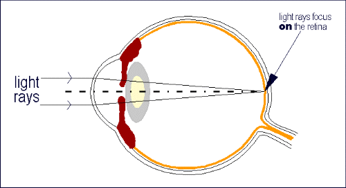
In hyperopia the distance between the cornea and the retina is smaller than normal. This results in a parallel beam which enters the eye to strike the retina before it is focused.
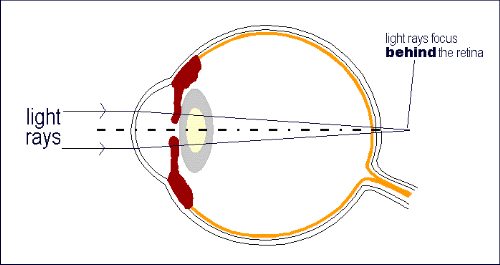
The causes of hyperopia are typically genetic and involve an eye that is too short or a cornea that is too flat, so that images focus at a point behind the retina. People with hyperopia can usually see distant objects well, but have trouble focusing on nearby objects. The activation of the adjustment mechanism can eliminate a large percentage of hyperopia.
In childhood there is no loss of visual acuity due to hyperopia. Children with hyperopia develop disorders in binocular vision, a tendency for convergent strabismus and kopiopia..
Astigmatism
Ophthalmic astigmatism is a refraction error of the eye in which there is a difference in degree of refraction in different meridians of the cornea. It is typically characterized by an aspherical, non-figure of revolution cornea in which the corneal profile slope and refractive power in one meridian is less than that of the perpendicular axis.
Astigmatism causes difficulties in seeing fine detail and is quite common. Studies have shown that about one in three people suffers from it. The prevalence of astigmatism increases with age. Although a person may not notice mild astigmatism, higher amounts of astigmatism may cause blurry vision, squinting, asthenopia, fatigue, or headaches.
Astigmatism can be often corrected by glasses with a lens that has different radii of curvature in different planes (a cylindrical lens), contact lenses, or refractive surgery.
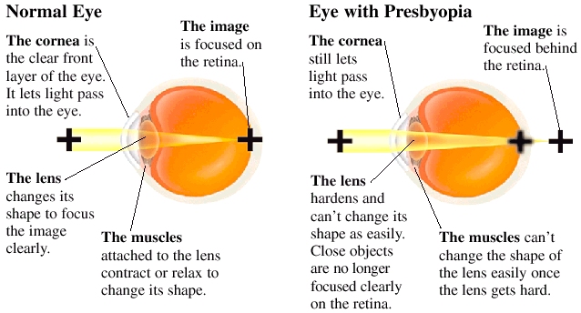
Presbyopia
Presbyopia occurs at the age of 40 years and its main symptom is reduced near vision. Difficulty in reading without glasses at 35-40 cm and fatigue after a short period of close work are present. Normally the lens is flexible enough to change its shape when focusing at close objects. Loss of its flexibility and elasticity known as loss of the eye’s adjustment mechanism results in presbyopia. In recent years VEIC is a leading Institute in the research for the development of effective and permanent surgical treatment for presbyopia.